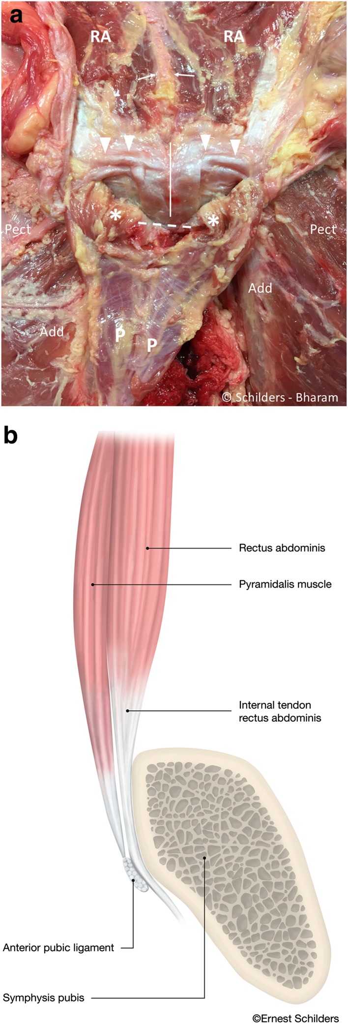
Fig. 6.
a Male cadaver, same specimen as Fig. 4. The pyramidalis muscle is sharply detached from the linea alba (white arrows) and folded distally. The internal tendons of the rectus abdominis run anterior to the symphyseal joint (white line), and the external tendon inserts on the superior lateral edge of the pubis (arrowheads). There is a thin aponeurosis on the posterior side of the pyramidalis muscle (P) with transverse fibre orientation. This aponeurosis does not cover the pyramidalis muscle anterior to the pubic bone (asterisk). Pect pectineus muscle. Add adductor longus muscle. A linea alba. Dashed line region of the anterior pubic ligament. RA rectus abdominis muscle. b Sagittal midline drawing demonstrating the anatomical relationship of the anterior pubic ligament (arrowhead), the pyramidalis muscle (P) and the internal tendon of the rectus abdominis (RA) at the level of the symphysis pubis. The internal tendon of the rectus abdominis receives contributions from left and right sides giving the tendon a Y-shape (arrow). The pyramidalis muscle inserts on the anterior pubic ligament, which spans the symphyseal joint and acts like a pulley through its position anterior to the internal tendon of the rectus abdominis muscle

