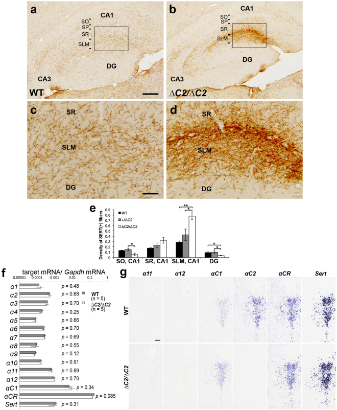
Figure 3.
αC2 protein is required for normal serotonergic projections. (a–d) Serotonergic axons in the hippocampus of WT (a,c) and Pcdha ∆C2/∆C2 (b,d) mice were detected with an anti-SERT antibody. Panels (c) and (d) are higher-magnification images of the boxed areas in (a) and (b), respectively. SP, stratum pyramidale. (e) Quantification of SERT(+) fibers in the hippocampus of WT (n = 4), Pcdha +/∆C2 (n = 3), and Pcdha ∆C2/∆C2 (n = 4) mice. *p < 0.05, **p < 0.01. Mean ± SEM. (f) Quantitative RT-PCR for the Pcdha and Sert genes. There was no significant difference between WT and Pcdha ∆C2/∆C2 KO mice. Mean ± SEM. (g) Expression analysis by in situ hybridization using probes for α11, α12, αC1, αC2, αCR, and Sert in adjacent coronal sections of the dorsal raphe nucleus of WT and Pcdha ∆C2/∆CR mice. Scale bars: (a,b), 200 µm; (c,d) 50 µm; 200 μm (g).

