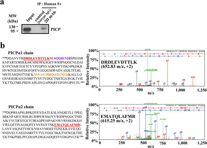
Figure 4.
Specific binding of 2D mAb to native PICP. (a) Western blot analysis of the anti-PICP 2D mAb immunoprecipitation (IP) for culture supernatants of primary dermal fibroblast cells. The corresponding full-length blots are shown in Supplementary Fig. 6. (b) Identification of PICP protein in the immunoprecipitated gel band by LC/MS. The three different peptides and one peptide identified from the PICPα1 and PICPα2 chains, respectively, were indicated in distinct colors in the amino acid sequence. The left panels show the representative MS/MS spectrum for the identified peptides of DRDLEVDTTLK (652.83 m/z, +2) from PICPα1 (top) and EMATQLAFMR (615.28 m/z, +2) from PICPα2 (bottom), underlined in each sequence.

