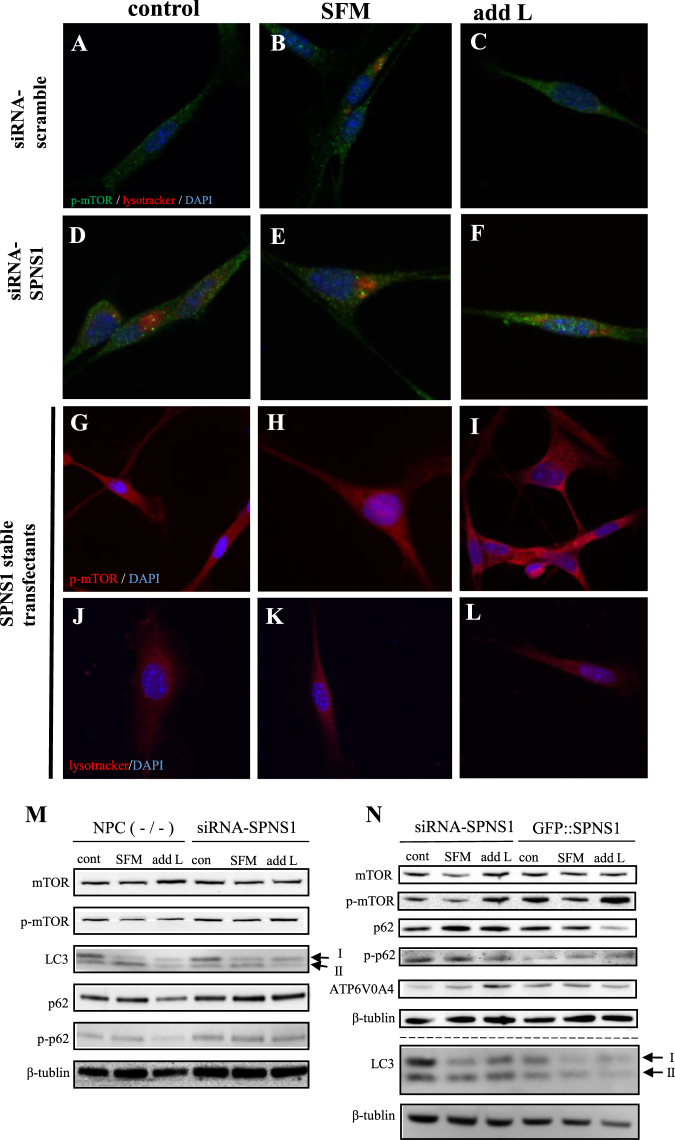
Figure 2.
Effects of SPNS1 knockdown and overexpression on autophagy defects of serum-deprived NPC (−/−) cells with or without L-leucine supplementation. (A–F) Localization of phosphorylated mTOR (p-mTOR) (green) and lysotracker (red) signals in NPC (−/−) cells without (A–C) or with (D–F) SPNS1 knockdown kept in a normal culture medium (control: A,D), serum-free-medium (SFM: B,E) or L-leucine supplemented serum-free medium (add L: C,F). (G–I) Localization of phosphorylated mTOR (p-mTOR) (red) in NPC (−/−) cells with SPNS1 stable expression kept in a normal culture medium (control: G), serum-free-medium (SFM: H) or L-leucine supplemented serum-free medium (add L: I). (J–L) Lysotracker (red) signals in NPC (−/−) cells with SPNS1 stable expression kept in a normal culture medium (control: J), serum-free-medium (SFM: K) or L-leucine supplemented serum-free medium (add L: L). (M) Western blot analysis of the indicated proteins in NPC (−/−) cells without (left) or with (right) SPNS1 knockdown (via siRNA-SPNS1). Cells were kept in a normal culture medium (con), serum-free medium (SFM) or serum-free medium supplemented with L-leucine (add L). The arrows indicate two forms of LC3 protein (LC3I and LC3II). (N) Western blot analysis of the indicated proteins in NPC (−/−) cells with SPNS1 knockdown (siRNA-SPNS1) or NPC (−/−) cells with stable SPNS1 expression (GFPSPNS1). Cells were kept in a normal culture medium (con), serum-free medium (SFM) or serum-free medium supplemented with L-leucine (add L). Arrows indicate two forms of LC3 protein (LC3I and LC3II).

