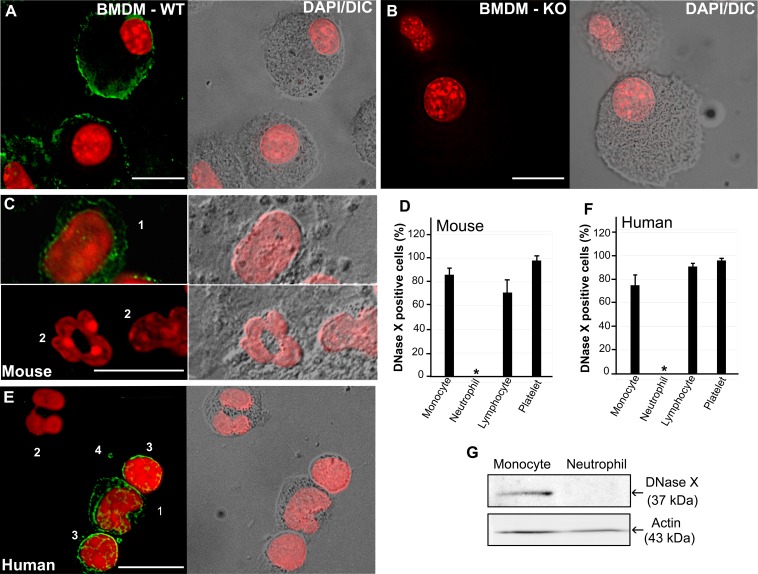
FIG 6 .
Human and mouse peripheral blood neutrophils do not express DNase X. Immunofluorescence labeling (A to C, E) and quantification (D, F) of WT and DNase X−/− BMDMs and human and WT mouse PBLs with antibodies against DNase X is shown. WT BMDMs (A), DNase X−/− (KO) BMDMs (B), WT mouse PBLs (C and D), and human peripheral blood leucocytes (E and F) were used. Cells: 1, monocyte; 2, neutrophil; 3, lymphocyte; 4, platelet. DeltaVision deconvolution fluorescence microscopy/differential interference contrast microscopy was used. Scale bars, 10 µm. Nuclei were stained with DAPI (pseudocolored red). (D and F) DNase X-expressing cells were scored to analyze a total of 100 of each type of cell from three individuals. Results are presented as the mean ± the standard deviation of three individuals and were compared by analysis of variance; *, P < 0.05. (G) Western blot analysis of human peripheral blood-derived monocytes and neutrophils with anti-DNase X and anti-actin antibodies.

