FIG 3 .
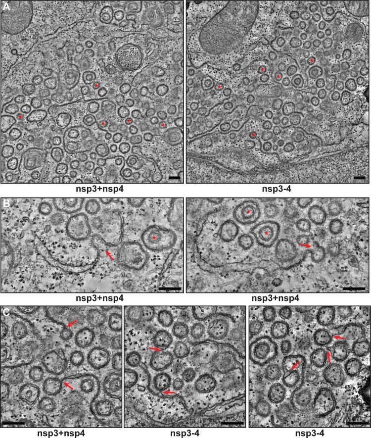
MERS-CoV nsp3 and nsp4 induce the formation of DMVs that are organized in an RVN. HuH-7 cells were cotransfected with constructs expressing nsp3 and nsp4 or the nsp3-4 precursor and fixed for ET analysis. (A) Overviews of reconstructed tomograms (available as Movies S1 and S2, respectively) for both conditions. Some of the fully reconstructed closed DMVs are indicated with red asterisks. (B) Zippered ER curving into putative intermediates during DMV biogenesis (indicated with red arrows) is shown. Two DMVs that are enclosed within other DMVs are indicated with red asterisks. (C) Examples of connections between DMVs and (zippered) ER (indicated with red arrows). All the images are virtual 5-nm-thick slices from the reconstructed tomograms. Bars, 250 nm.

