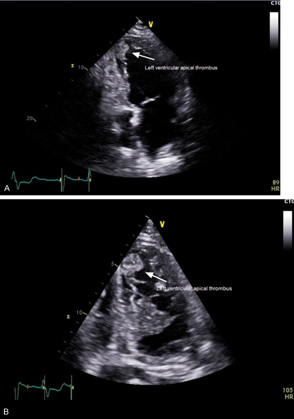
Figure 1.

A. This is an apical view of the left atrium and left ventricle and the presence of a left ventricular thrombus in the apex. B. This is an enlarged apical view of the left ventricle again demonstrating the left ventricular apical thrombus. Due to the significant size of the thrombus, there was no need to confirm this finding with the use of contrast.
