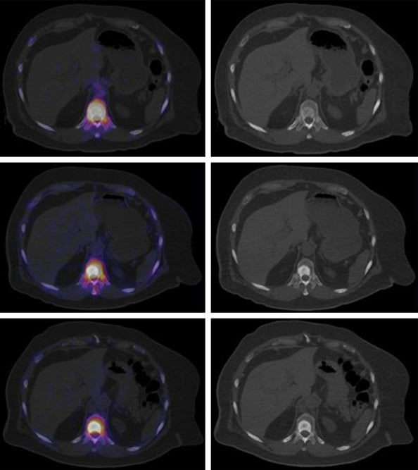
Figure 5.

Top. Fused axial slice of baseline 18F-NaF-PET/CT and high radionuclide uptake (left), axial slice showed blastic lesion in vertebral body (right). Middle. Decrease focal uptake in blastic lesion (left), important increase of sclerotic component, after 3 cycles of 223Ra is seen. Lower. decrease focal uptake in blastic lesion (left), note the increase of sclerotic component, after 6 cycles of 223Ra plus ADT.
