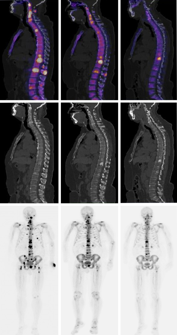
Figure 6.

Top Left. Fused sagittal slice of baseline 18F-NaF-PET/CT and high radionuclide uptake in multiple vertebral bodies. Top middle. Fused sagittal slice 18F-NaF-PET/CT after 3 cycles of 223Ra. Top Right. Fused sagittal slice 18F-NaF-PET/CT after 6 cycles of 223Ra plus ADT, note the important osteoblastic activity. Middle. Sagittal slices corresponding only a CT baseline, CT after 3 cycles of 223Ra and CT after 6 cycles of 223Ra plus ADT, note the increased sclerotic changes. Lower Left. multiple sites of focal high radionuclide uptake that result in metastatic bone disease predominantly blastic type axial skeleton in the MIP 18F-NaF-PET. Lower middle. MIP 18F-NaF PET after 3 cycles of 223Ra. Lower Right. MIP 18F-NaF-PET after 6 cycles of 223Ra plus ADT, note the important decreased of osteoblastic activity (patient 4).
