
Figure 1.
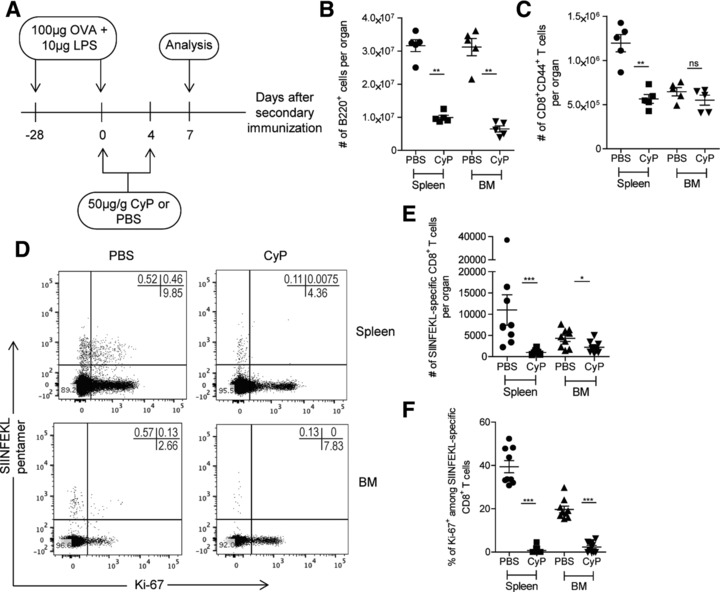
Proliferating CD8+ T lymphocytes are eliminated by CyP in acute immune reactions. (A) Experimental setup: CyP was applied on days 0 and 4 of a secondary immune response to Ova. Numbers of specific CD8+ T cells were determined in spleen and BM on day 7 of the secondary immune response. (B) Absolute numbers of B220+ cells in spleen and BM upon administration of CyP or vehicle. (C) Absolute numbers of CD8+CD44+ T cells in spleen and BM upon administration of CyP or vehicle. (D) Representative dot plots of SIINFEKL‐pentamer versus Ki‐67 gated on CD4−CD8+CD44+ viable cells. (E) Absolute numbers of SIINFEKL‐specific CD8+ T cells in spleen and BM upon administration of CyP or vehicle. (F) Frequencies of Ki‐67+ among SIINFEKL‐specific CD8+ T cells in spleen and BM upon administration of CyP or vehicle. Data in (B) and (C) are representative of two independent experiments, each with four to five mice per group. Data in (E) and (F) represent pooled results from two independent experiments, each with four to five mice per group. Data are presented as mean ± SEM. *p < 0.05, ** p < 0.01, *** p < 0.001, as determined by two‐tailed Student's t test.