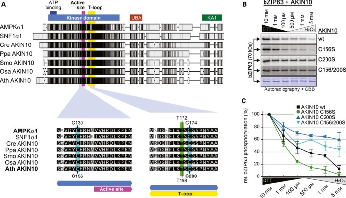
Figure 2.

Redox sensitivity of AKIN10 partially depends on a conserved cysteine residue in its T‐loop. (A) Protein sequence alignment of AKIN10 orthologues in human (AMPKα1), yeast (SNF1α1) and plants. Chlamydomonas reinhardtii (Cre AKIN10), Physcomitrella patens (Ppa AKIN10), Selaginella moellendorffii (Smo AKIN10), Oryza sativa (Osa AKIN10) and Arabidopsis thaliana (Ath AKIN10) are shown as representatives of Chlorophytes, Bryophytes, Lycophytes, Monocots and Eudicots respectively. Sequences were aligned in Geneious using the default ClustalW settings. On the top, the complete sequence alignment is shown. The shaded boxes indicate the sequence conservation. The positions of the kinase domain (blue), the ATP‐binding site (dark blue), the active site (purple), the T‐loop (yellow), the ubiquitin‐associated domain (UBA, red) and the kinase‐associated 1 motif (KA1, green) are indicated. On the bottom, neighbouring sequences of C130 and C174 in AMPK are shown (highlighted in cyan). The crucial threonine in the T‐loop is highlighted in green. The shading indicates the degree of sequence similarity (black: 100%, dark grey: 80–100%, light grey: 60–80%, white: < 60%). An extended alignment of plant AKIN10 orthologues, including 52 different plant species, can be found in Fig. S2. (B) In vitro phosphorylation of bZIP63 by different AKIN10 variants. GST‐bZIP63 (bZIP63) was incubated with GST‐AKIN10 (wt) or three different cysteine to serine mutants (C156S, C200S and C156/200S) in kinase buffer containing 32P γATP and different concentrations of either DTT or H2O2. The proteins were separated by SDS/PAGE and phosphorylated proteins were detected by subsequent autoradiography. A representative coomassie brilliant blue‐stained gel is shown below. Relative quantification of three independent assays is shown in Fig. 2C. (C) Relative quantification of GST‐bZIP63 phosphorylation by GST‐AKIN10 (black squares) and its cysteine to serine variants (C156S, green circles; C200S, dark blue triangles; C156/200S, light blue triangles). The autoradiography values obtained from kinase reactions containing 10 mm DTT (highest kinase activity in all instances) were defined as 100%. The values are the mean ± standard deviation of three independent experiments.
