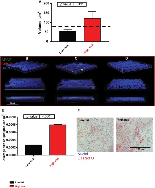
Figure 4.

Drüsen‐like deposits form in high‐risk induced pluripotent stem cell (iPSC)‐retinal pigment epithelium (RPE). (A): Volume of apolipoprotein E (APOE) and C5b‐9 coexpressing deposits in low‐ and high‐risk iPSC‐RPE, dashed black line represents clinically significant drüsen size. High‐risk donor iPSC‐RPE cells accumulate larger deposits than low‐risk, n = 3. (B): Low‐risk iPSC‐RPE example of F116; blue, 4′,6‐diamidino‐2‐phenylindole (DAPI); red, C5b‐9; and green, APOE. Scale bar = 50 µm. (C): High‐risk iPSC‐RPE example of F181; blue, DAPI; red, C5b‐9; and green, APOE. Scale bar = 50 µm. (D): Secondary antibody only control; blue, DAPI, red, C5b‐9; and green, APOE. Scale bar = 50 µm. (E): Oil red O staining, high‐risk donor iPSC‐RPE contained larger lipid globules than low‐risk donors. (F): Examples of low‐ and high‐risk donor oil red O staining. Scale bar = 200 µm. Abbreviations: APOE, apolipoprotein E; DAPI, 4′,6‐diamidino‐2‐phenylindole.
