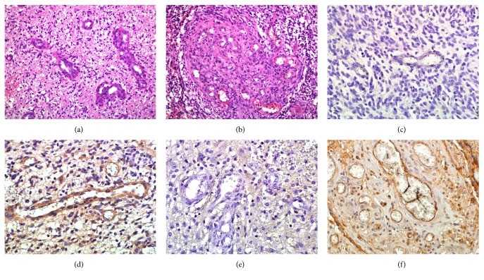
Figure 1.
Blood vessels with various characteristics in glioblastoma (GB). The proportion of microvascular proliferation varied from low-medium to high. (a) Example of a case with sparsely located small microvascular proliferation (H&E, ×20). (b) Example of abundant large size microvascular tufts (H&E, ×20). (c) Immunohistochemical staining with anti-CD-133 antibody revealed portion of capillary blood vessels with CD133-negative staining (×40). (d) GB blood vessels with CD133-positive vascular staining (×40). (e) GB blood vessels with weak immunohistochemical staining for ICAM-1 (×40). (f) GB blood vessels with strong immunohistochemical staining for ICAM-1 (×40).

