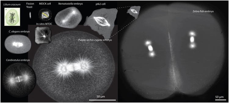
Figure 1. Precision of centering the mitotic spindles during metaphase in various eukaryotic cells and in-vitro systems.

Clockwise from the upper left: Mitotic cell drawn by W. Fleming in 1882 [108] (Not at scale). Fission yeast cell (kindly provided by Prof. Iva Tolic). A HeLa cell with fluorescently labeled (α-tubulin antibody) microtubules, DNA and actin (Prof. Linda Wordeman). Single cell from a Nematostella embryo (32-cell stage) fluorescently labeled with EB1-GFP (Dr. Katerina Ragkousi – Gibson Lab). Sea urchin zygote with fluorescently labelled microtubules (Prof. Victoria Foe). Single-cell Cerebratulus marginatus zygote with fluorescently labelled microtubules and DNA (Prof. George von Dassow). C. elegans zygote with fluorescently labelled microtubules (β-tubulin:GFP). Fluorescently labelled microtubules growing from a fluorescently labelled bead inside a squared-shaped chamber (Prof. Marleen Dogterom). Compressed PTK2 cell labeled with GFP-α-tubulin (Joshua Guild – Dumond Lab). Four–cell zebrafish embryo with fluorescently labelled microtubules (Elisa Rieckhoff – Brugues Lab). Notice that the zebrafish embryo has a different scale bar.
