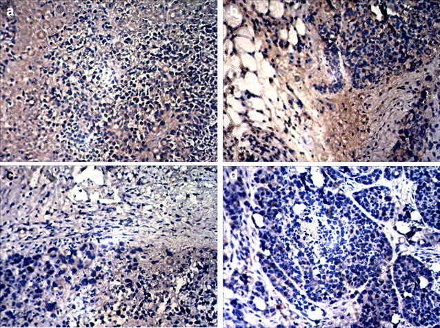
Fig. 8.

Immunohistochemical staining of CD34 in residual tumor tissues (×200). The number of positively stained areas of tumor tissues in each group varied. (a saline; b 5-FU-loaded nanobubbles; c 5-FU-loaded nanobubbles with non-low-frequency ultrasound; d 5-FU-loaded nanobubbles with low-frequency ultrasound)
