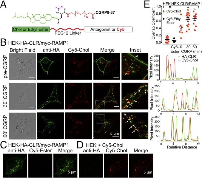
Fig. 3.
Tripartite probes. (A) Probe structure. (B–D) Confocal images of live HEK cells. (B and C) HEK-HA-CLR/myc-RAMP1 cells were incubated with Cy5-Chol (B) or Cy5-Ethyl Ester (C). CLR was labeled with HA-Alexa488 antibody. Cells were stimulated with CGRP (50 nM, 30 or 60 min) to induce endocytosis. Insets (white boxes) show magnified regions and colocalization (arrows). Traces (Left) show relative overlap of pixel intensities for HA-CLR and Cy5-Chol along dashed lines. (D) Untransfected HEK cells incubated with Cy5-Chol. (E) Overlap coefficient for HA-CLR and Cy5-Chol or Cy5-Ethyl Ester. n = 6–14 cells, n = 4 experiments. **P < 0.01 to 0 min. ANOVA, Dunnett’s test.

