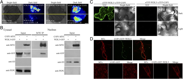
Fig. 3.
Interaction of COP1 with WDL3 at microtubules. (A) WDL3 did not interact with COP1-△R revealed by the firefly LCI assays in tobacco leaves in the dark. HYH was used as a positive control and exhibited interaction with COP1 or COP1-△R under the same conditions. (Scale bar: 1 cm.) (B) Coimmunoprecipitation of cytoplasmic COP1 and WDL3 extracted from tobacco leaves expressing 35S:COP1-MYC and 35S:WDL3-GFP. Anti-MYC-Agarose Affinity Gel was used for immunoprecipitation, and the precipitated proteins were analyzed by immunoblots using an anti-GFP antibody. PEPC and H3 were used as cytosolic and nuclear markers, respectively. (C) BiFC assays showing interaction between COP1 and WDL3 in tobacco leaves in the dark. Negative controls used were nYFP-WDL5+cYFP-COP1. (Scale bar: 20 μm.) (D) Confocal microscopy images of microtubules polymerized from rhodamine-labeled tubulin (20 μM) incubated in the presence or absence of 3 μM GST-WDL3 or GST-WDL5 with COP1-GFP fusion proteins for 20 min. (Scale bar: 10 μm.)

