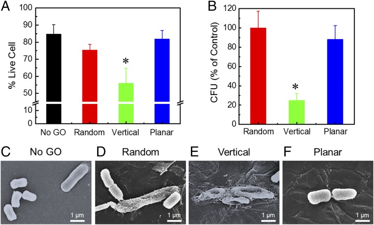
Fig. 4.
Antibacterial activity of GO composite films with different orientations of GO nanosheets. (A) Cell viability of deposited E. coli cells after 3 h of contact with surfaces with aligned GO nanosheets, determined by live/dead fluorescent staining assay. Values marked with an asterisk (*) are significantly different from the value of No-GO sample (n = 3; Student’s t test, P < 0.05). (B) Relative number of viable E. coli cells after 3 h of contact with surfaces with aligned GO, determined by cfu plate counting and normalized to the results of the Random-GO surface. The No-GO surface was not used as a control due to decreased bacterial adhesion stemming from the smooth and hydrophilic nature of the poly-HEMA surface. Values marked with an asterisk (*) are significantly different from the value of Random sample (n = 3; Student’s t test, P < 0.05). Representative SEM micrographs of E. coli cells on polymer films with No GO (C) and Random (D), Vertical (E), and Planar (F) GO nanosheets. The bacteria on the No-GO film showed intact cell morphology. Among the GO composite films, the cells on the Random-GO and Planar-GO films largely retained their morphological integrity, whereas the cells on the Vertical-GO films became flattened and wrinkled, suggesting loss of viability and possible damage to the cell membrane.

