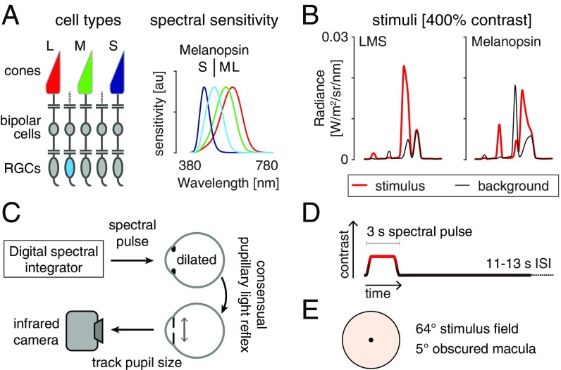
Fig. 1.
Overview and experimental design. (A, Left) The L, M, and S cones, and melanopsin-containing ipRGCs, mediate visual function at daytime light levels. (A, Right) The spectral sensitivities of these photoreceptor classes. (B) Stimulus spectra. Changes between a background (black) and stimulus (red) spectra targeted a given photoreceptor channel. The 400% contrast stimuli are shown. (B, Left) Spectra targeting the L, M, and S cones and thus the postreceptoral luminance channel. The nominal melanopic contrast for this modulation was zero. (B, Right) The corresponding spectra for stimuli targeting melanopsin. The nominal L-, M-, and S-cone contrast of this stimulus was zero. (C) During fMRI scanning, subjects viewed pulsed spectral modulations, produced by a digital spectral integrator, with their pharmacologically dilated right eye. The consensual pupil response of the left eye was recorded in some experiments. (D) Multiple 3-s, pulsed spectral modulations were presented, windowed by a 500-ms half-cosine at onset and offset, and followed by an 11- to 13-s interstimulus interval (ISI). A given experiment presented either a single contrast level, or multiple contrast levels in a counterbalanced order. (E) Spectra were presented on a uniform field of 64° (visual angle) diameter. Subjects fixated the center of a 5° masked region, minimizing macular stimulation. The stimulus spectra had a light-orange hue.

