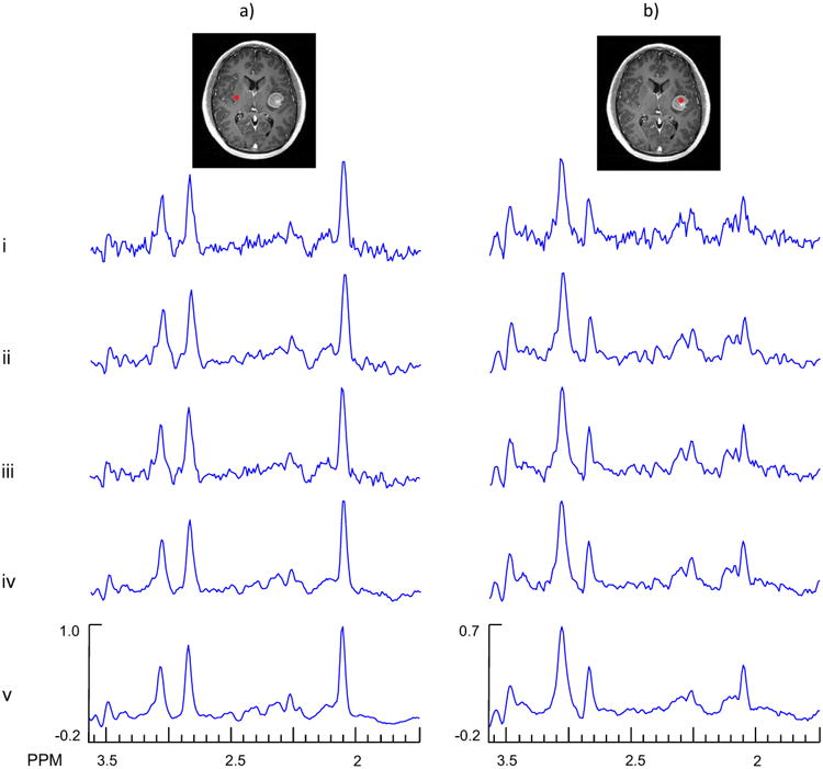
Fig. 6.

Example spectra from the denoising of an in vivo MRSI study of a subject with brain tumor. In column a) are shown results for a voxel selected from a normal appearing white matter region (voxel 1) and in column b) are shown results for a voxel selected in the tumor region (voxel 2). The rows show results for the original data (i), Gaussian denoising (ii), PCA denoising using the first 119 PCs (iii), SSPC denoising with a threshold of 1E-5 (the algorithm found 119 PCs for this threshold) (iv), and SSPC denoising with a threshold of 1E-10 (the algorithm found 56 PCs for this threshold) (v).
