
Figure 4. Smurf1 Mediates K48-Linked, But Not K63-Linked, Polyubiquitination of M. tuberculosis and L. monocytogenes.
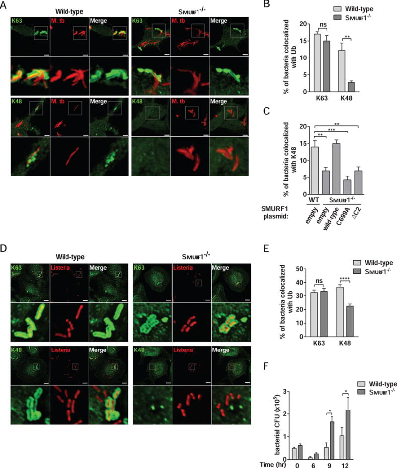
(A and B) Photomicrographs (A) and quantitation (B) of the colocalization of mCherry-Mtb and K63 or K48 polyubiquitin in wild-type or Smurf1−/− BMDMs 15 hr after infection. Scale bars, 3 μm. Bars are mean ± SEM for quadruplicate samples (at least 100 bacteria analyzed per sample) from a representative experiment. **P<0.01; ns: non-significant; t-test.
(C) Rescue of mycobacterial colocalization with K48-linked polyubiquitin by wild-type but not mutant forms of SMURF1 in Smurf1−/− BMDMs. Wild-type BMDMs were transduced with empty lentivirus and Smurf1−/− BMDMs were transduced as described in Figure 3B, and infected with mCherry-Mtb for 15 hr. Bars are mean ± SEM for quadruplicate samples (at least 100 bacteria analyzed per sample) from a representative experiment. **P<0.01; ***P<0.001; one-way ANOVA.
(D and E) Photomicrographs (D) and quantitation (E) of the colocalization of RFP-expressing L. monocytogenes ΔActA mutant and K63 or K48 polyubiquitin in wild-type or Smurf1−/− BMDMs 2 hr after infection. Scale bars, 5 μm. Bars are mean ± SEM for quadruplicate samples (at least 100 bacteria analyzed per sample) from a representative experiment. ****P<0.0001; ns: non-significant; t-test.
(F) L. monocytogenes growth in wild-type or Smurf1−/− BMDMs. Infected cells were lysed at the indicated time-points and L. monocytogenes growth was determined by counting CFU. Bars are mean ± SEM for quadruplicate samples. *P<0.05 for indicated comparison; two-way ANOVA. For B, C, E, and F, similar results were observed in at least three independent experiments.
See also Figure S2.