
Figure 5. Smurf1 Cooperates With Parkin to Trigger Ubiquitin-Dependent Selective Autophagy of M. tuberculosis.
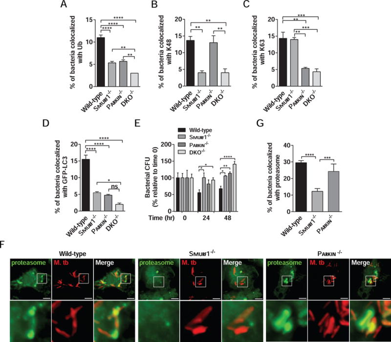
(A–D) Quantitation of the colocalization of mCherry-Mtb and polyubiquitin (A), K48 (B), K63 (C) or GFP-LC3 (D) in wild-type, Smurf1−/−, Parkin−/− or Smurf1−/−Parkin−/− double knockout (DKO−/−) BMDMs 15 hr after infection. Bars are mean ± SEM for quadruplicate samples (at least 100 bacteria analyzed per sample) from a representative experiment. *P<0.05; **P< 0.01; ***P<0.001; ****P<0.0001 for indicated comparison; ns: non-significant; one-way ANOVA.
(E) Mtb growth in wild-type, Smurf1−/−, Parkin−/− or DKO−/− BMDMs. Infected cells were lysed at the indicated time-points and Mtb growth was determined by counting CFU. Bars are mean ± SEM of quadruplicate samples; each sample was normalized to day 0. *P<0.05; **P< 0.01; ***P<0.001; ****P<0.0001 for indicated comparison; two-way ANOVA.
(F and G) Photomicrographs (F) and quantitation (G) of the colocalization of mCherry-Mtb and the proteasome 15 hr after infection of BMDMs from wild-type, Smurf1−/− or Parkin−/− mice. Insets (lower panels) in (F) show representative mycobacteria that would be considered colocalized with the proteasome in wild-type or Parkin−/− BMDMs or not colocalized with the proteasome in Smurf1−/− BMDMs. In (G), bars are mean ± SEM of quadruplicate samples (100 bacteria evaluated per sample per genotype) from a representative experiment. Scale bars, 5 μm. ***P<0.001; ****P<0.0001 for indicated comparison; one-way ANOVA. For For A–E and F, similar results were observed in at least three independent experiments.
See also Figure S3.