
Figure 2.
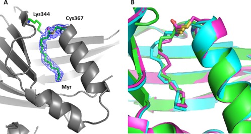
X‐ray structure of myristoylated human hTEAD4217–434. The coordinates have been deposited in the PDB databank (PDB access code = 5OAQ). For clarity, the bound YAP60–100 peptide is not displayed (see Supporting Information Fig. S1). (A) Acylation site. TEAD4 is represented by gray ribbons. hTEAD4 Cys367, Lys344, and the bound myristate moiety are indicated. The electron density (2Fo‐Fc map contoured at 1σ) for Cys367 and the bound myristate are shown (blue mesh). (B) The structure of hTEAD4217–434 (green), hTEAD2217–447 (pdb 5EMV, cyan) and hTEAD3216–435 (pdb 5EMW, purple) have been superimposed. The modified cysteines (hTEAD4 Cys367, hTEAD2 Cys380, hTEAD3 Cys371) and the myristate (TEAD4) or the palmitate (TEAD2/3) moieties are represented. The figures were generated with PyMol (Schrödinger LLC).