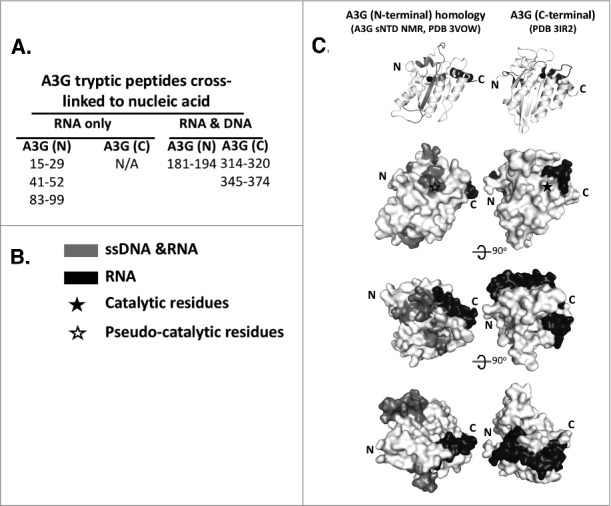
Figure 3.

RNA and ssDNA Binding Surfaces on A3G. (A) Tryptic peptides of A3G bound to RNA or ssDNA were identified by mass spectroscopy following cross linking of native and full length A3G to short nucleic acids. (B) Peptides that only bound RNA (black) or bound to both RNA and ssDNA (gray) were mapped relative to the C-terminal ZDD catalytic domain and the N-terminal ZDD catalytically inactive pseudo-catalytic domain. (C) Grey scale coded RNA binding peptides and RNA and ssDNA binding peptides were mapped onto the NMR structure for the N-terminal ZCC and the crystal structure of the C-terminal ZDD of A3G shown as a ribbon diagram (top) and progressively rotated (top to bottom) space filling models. Black star and open star mark the location of the the catalytic and pseudo-catalytic ZDD, respectively. Reproduced with permission from reference 31.
