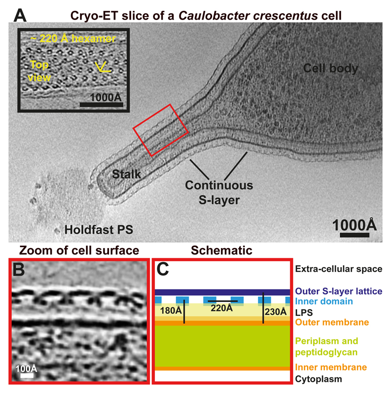
Figure 1. Arrangement of the Caulobacter crescentus S-layer on cells and stalks.
(A) A tomographic slice of a CB15 C. crescentus cell. The S-layer is continuous between the cell body and the stalk. Inset – a magnified tomographic slice through a S-layer of a stalk. A hexameric lattice with a ~220 Å spacing is seen (see also Movie S1). (B) A magnified tomographic slice of a side view of the cell surface. The S-layer is arranged in two layers and is seen ~180 Å away from the outer membrane of the cell. The outer S-layer lattice is highly inter-connected while the inner domains are ~220 Å apart from each other. (C) A schematic representation of the cell surface.

