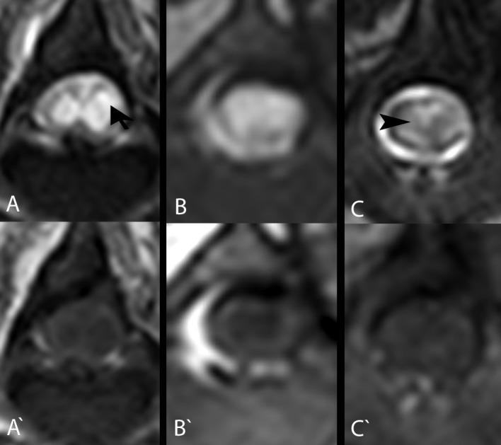
Fig 2. Measurement of the length, maximal cross-sectional area (mCSA) of intramedullary lesions/cavitations and spinal cord diameter at the lesion epicentre.

The length of intramedullary lesions/cavitations was estimated by measuring the length of the intramedullary lesions/cavitations in T2W MR images and calculating the ratio to the length of the L2 vertebra (A). Maximal CSA of spinal cord lesions/cavitations was measured by measuring the area of the spinal cord lesion/cavitation in the T2W MR images (B) and dividing it by the area of the spinal cord at the same level (B‘), which led to a percentage of the damaged spinal cord. Spinal cord diameter was measured in T2W MRI at the lesion epicentre (C ‘) and at the level of the normal spinal cord cranially (C) and caudally (C ‘‘). The difference of diameters led to the estimated reduction of spinal cord diameter.
