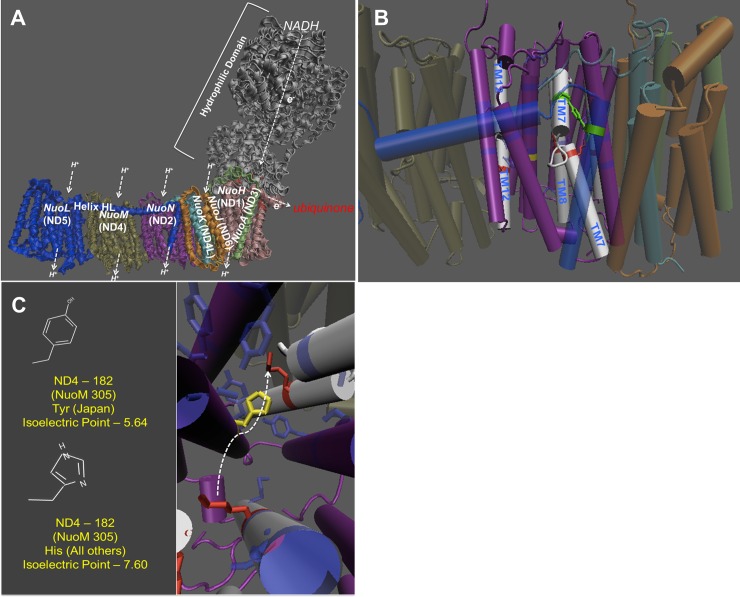
Fig 1. Complex I structure prediction of Japan mtDNA.
(A) The ND2 region corresponds to the bacterial subunit NuoN. (B) The structure of the proton pump. The dark blue residues that are transparently shaded make up the proton channel. (C) The location of the variant and their isoelectric point. The yellow is the amino acid that is changed in the Japan line. The arrow represents the movement of the TM7 domain.

