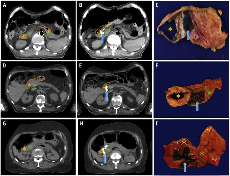
Fig. 2.

Computed tomography scans before and after hydrogel spacer injection between the head of the pancreas and duodenum, with postinjection gross histologic specimens confirming location of the spacer. (A-C) Gel placed using laparotomy. Gel placed endoscopically in (D-F) EUS cadaveric specimen 1 and (G-I) EUS cadaveric specimen 2. Duodenal lumen (orange outline and asterisk), hydrogel spacer (blue arrow), and head of the pancreas (green asterisk) are denoted.
