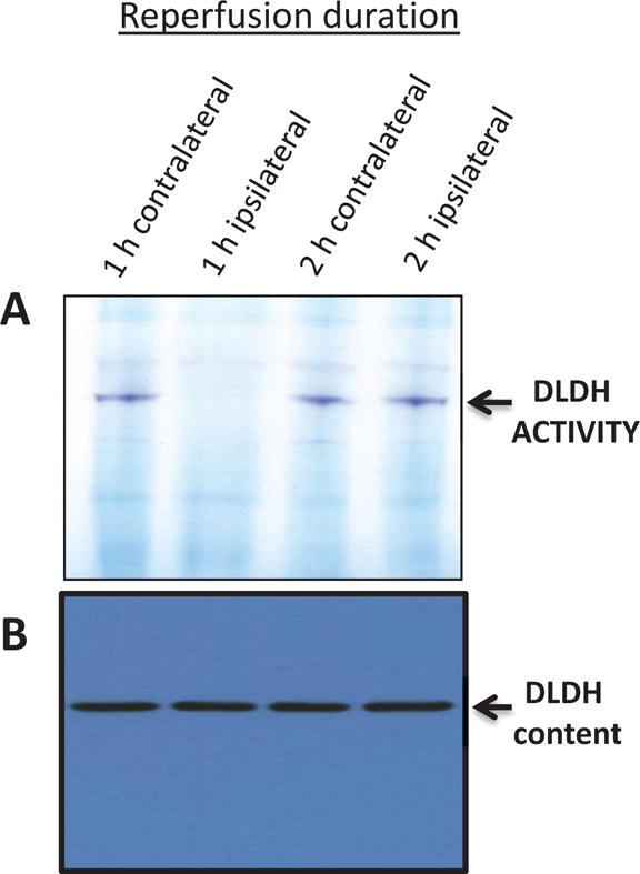
Fig. 9.

Measurement of DLDH activity (A) and protein content (B) after ischemia reperfusion. Animals were not treated with MICA. One h ischemia was followed by 1 or 2 h reperfusion. This was further followed by brain mitochondria isolation for gel analysis. DLDH activity was measured by in-gel BN-PAGE assay (upper panel) as described in the text, and DLDH protein content was measured by anti-DLDH Western blot assay (lower panel). Shown are contralateral samples and ipsilateral samples after 1 and 2 h reperfusion. As can be seen in the upper panel, DLDH activity was lost after 1 h reperfusion but recovered after 2 h reperfusion, but protein content did not change.
