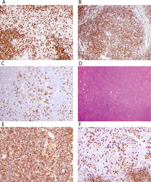
FIGURE 2.

Examples of variant histology. (A) Fan type C histology shows large cells labeling with anti-CD20 in extranodular areas. (B) Small lymphocytes in nodules of this case label with anti-CD3, identifying type D NLPHL. (C) CD20 highlights LP cells in the CD20-negative T-cell background of nodules in type D. (D) This hematoxylin and eosin (H&E) stain shows an apparently diffuse infiltrate in a case with type E morphology. (E) This case has >25% with diffuse small lymphocytes labeling with anti-CD20 as diffuse B-cell type D. This case is not as “moth-eaten” as usually described. (F) This case labeled with anti-CD20 shows type C with increased extranodular large B cells, merging into a diffuse type E pattern
