
FIGURE 4.
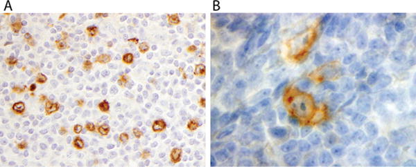
(A) Extranodular LP cells show cytoplasmic IgD staining in a background of small T cells in this case that includes score 2 types C and E, along with score 1 type A; (B) LP cells labels with anti-CD30
Official websites use .gov
A
.gov website belongs to an official
government organization in the United States.
Secure .gov websites use HTTPS
A lock (
) or https:// means you've safely
connected to the .gov website. Share sensitive
information only on official, secure websites.
(A) Extranodular LP cells show cytoplasmic IgD staining in a background of small T cells in this case that includes score 2 types C and E, along with score 1 type A; (B) LP cells labels with anti-CD30