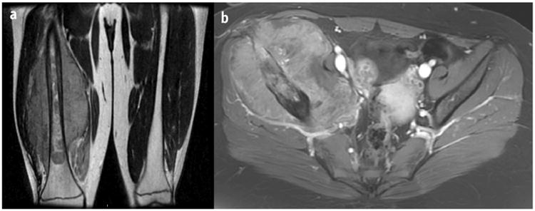
Fig. 2.

(a) Coronal T2 magnetic resonance image of a right lower extremity Ewing sarcoma tumor treated with radiation therapy. The tumor extended 30.0 cm along the right femur and was associated with a soft-tissue mass measuring 23.0 × 22.0 × 12.6 cm. Surgery would involve a non–limb-sparing approach. (b) Axial contrast T1 spoiled gradient magnetic resonance image of a right pelvis Ewing sarcoma tumor treated with radiation therapy. The tumor measured 15.0 × 13.2 × 9.3 cm and extended from the ilium to the superior pubic ramus. Surgery would require a hemipelvectomy.
