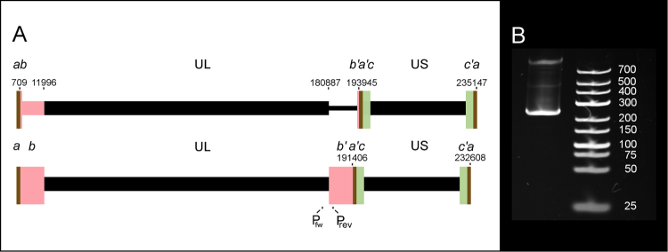
Figure 1.
Genomic rearrangement of the HCMV isolate used in this study. Panel A shows schematic representations of the original Towne strain virus above) and the isolate used in our experiments (below). The unique long (UL) and unique short (US) sequences are bracketed by repeat sequences (a–c), marked by coloured rectangles (brown, pink and green, respectively). In our isolate, the UL/b’ (180887–193945) region of the FJ616285 genome is substituted by the 710–11996 region. To confirm this rearrangement, primers have been designed to in the ends of the UL (Pfw) and b’ (Prev) regions 243 nt apart. Panel B shows the PCR product of approximately 250 nt. The original gel photo, from which Panel B was cropped is shown in Supplementary Fig. S1.

