Fig. 2.
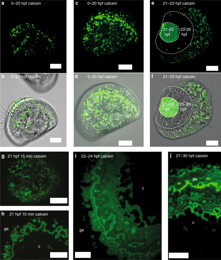
Calcein pulse-chase experiments and in vivo confocal microscopy from Exp 2. a, b Calcein fluorescence image and merged fluorescence and transmission images of larva cultured in calcein between 0–20 hpf, followed by development in filtered seawater (FSW) until 48 hpf, 1 μm thick section through body and shell to illustrate lack of calcein fluorescence in the shell. c, d Same animal, confocal projection through one entire shell valve and body to illustrate numerous calcein positive intracellular vesicles yet no fluorescence in the shell. e, f Confocal projection of calcein fluorescence of larva cultured in calcein FSW between 21–23 hpf. Shell material formed between 21–23 hpf is calcein labeled, shell material formed between 23–28 hpf is not. Calcein label of vesicles present at 28 hpf has apparently not been transferred into the shell. g, h 21 hpf animals stained with calcein for 15 min, then washed and cultured in FSW, calcein positive particles on periostracum g, shell growth bands in a slightly more advanced larva from the same fertilization h. i Confocal projection of shell calcein fluorescence of larva cultured in seawater with calcein pulse between 22–24 hpf. j Confocal projection of shell calcein fluorescence of larva cultured in seawater with calcein pulse between 27–30 hpf. c = centre of shell valve, ge = growing edge of shell valve. Scale bars: 20 µm a–f, 5 µm h, i, 10 µm j

