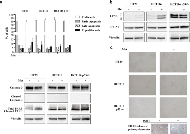
Figure 3.
Metformin (Met) does not induce apoptosis, autophagy and senescence in HT29, HCT116 and HCT116 p53−/− cells. (a) Graphic representation of the results obtained from Annexin V assay (upper panel) and western blot analysis of Caspase-3 and PARP activation after treatment with 5 mM Met for 72 h (lower panel). Vinculin was used as the loading control. (b) Immunoblotting determination of LC3B and BECN1 protein after treatment with 5 mM Met for 72 hours; Vinculin was used as the loading control. Full-size blots are shown in Supplementary Fig. S11. (c) β-galactosidase staining of untreated cells (−) and cells treated with Met for 72 hours (+). The images were acquired at 20x magnification. At the bottom, human primary thyrocytes carrying the ER:RAS vector were left untreated or treated with 200 nM 4-hydroxytamoxifen (4OHT) for seven days, and used as positive controls (magnification 40x). The data are representative of at least three independent experiments.

