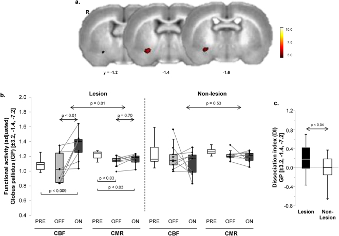
Figure 1.
Dissociation of vasomotor and metabolic response to levodopa in the unilateral 6-OHDA rat dyskinesia model. (a) Voxel-wise searches over the whole brain volume revealed a distinct region in which local vasomotor (CBF) and metabolic (CMR) changes were significantly dissociated in response to levodopa. This cluster (red), comprised of 49 contiguous voxels (1 voxel = 0.8 × 0.8 × 0.8 mm) located at the border of the striatum and the ventral globus pallidus (GP) of the dopaminergically denervated right (R) cerebral hemisphere, was significant at a voxel-level threshold of p < 0.001 corrected for extent at p < 0.05 (see text). (b) Box-and-whisker plots of CBF and CMR values in this region. Left: Significant CBF–CMR dissociation was seen in the lesioned hemisphere (F(1,8) = 10.85, p = 0.01, tracer × condition interaction effect; RMANOVA) with increased CBF (p < 0.01; post-hoc test) and no change in CMR (p = 0.70) in the OFF vs. ON condition. Right: Analogous changes were not seen in the contralateral non-lesioned hemisphere (p = 0.53). [Horizontal bars below box plots represent paired Student’s t-tests.] (c) Levodopa-mediated dissociation responses in this region were measured in the dopaminergically denervated and non-denervated hemispheres (see Methods). Dissociation responses were significantly greater on the 6-OHDA lesioned side relative to its non-lesionsed counterpart (p < 0.04; paired Student’s t-test).

