Figure 1.
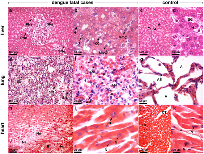
Histological injuries in the peripheral organs of dengue fatal cases (cases 1 and 2). Sections were stained with HE. (a) Hepatic parenchyma showing diffuse steatosis. The hepatocytes present swollen and irregular aspects that are classical cellular alterations of necrotic cells in periportal (PNe) and sinusoidal (SNe) areas. (b) Sinusoidal region showing hepatocytes (H), Kupffer cells (KC) and circulating macrophages (MϕC) with chromatin condensation and cytoplasm with bubble-like protuberances. (c) and (d) Liver sections of a non-dengue case presenting normal hepatic parenchyma (c) and hepatocytes (H) and sinusoidal capillaries (SC) presenting regular aspects of nuclei and cytoplasm (d). (e) Pulmonary parenchyma with relevant hemorrhagic areas, hypercellularity, septal thickening and septal necrosis (SN). (f) Region of septal thickening presenting numerous alveolar macrophages (AM) with loss of membrane integrity and altered heterochromatin. (g) Non-dengue Lung section showing normal alveolar septum (AS) structure and alveolar septal cells with regular characteristics (A - alveoli). (h) Extensive areas of necrosis (Ne), edema (E) and hemorrhage (He) in the cardiac parenchyma. (i) Cardiomyocytes presenting apoptotic nuclei (N) with heterochromatin damages, loss of membrane integrity and striations. (j) and (k) Heart sections of a non-dengue case showing (j) normal aspects of cardiac fibers (CF), conjunctive tissue (CT) and (k) cardiomyocytes with regular chromatin distribution (N - nucleus).

