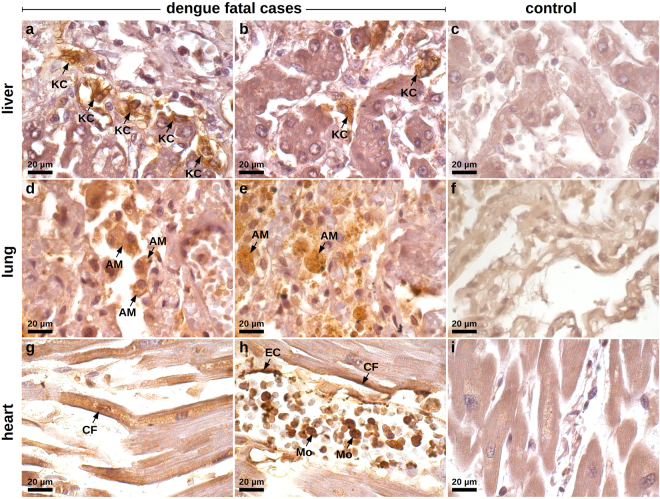
Figure 4.
Screening of HMGB1 expression in peripheral organs of dengue fatal cases by immunohistochemistry. (a) Hepatic parenchyma of samples collected from dengue case 1 showing hyperplasic and hypertrophic Kupffer cells (KC) with damaged cell membrane and HMGB1 expression within the cytoplasmic region. (b) Hepatic parenchyma exhibiting HMGB1 expression in the interior of vesicles of activated Kupffer cells (case 1). (c) Non-dengue case showing regular hepatic parenchyma and with basal HMGB1 expression. (d) Lung sections showing alveolar septal thickening region containing numerous alveolar macrophages (AM) expressing cytoplasmic HMGB1 (case 2). (e) Activated alveolar macrophages expressing HMGB1 inside cytoplasmic vesicles (case 2). (f) Liver sections from a non-dengue case presenting regular structure of alveolar septum with a marginal nuclear expression of HMGB1. (g) HMGB1 expression along the periphery of necrotic cardiac fibers (CF) (case 1). (h) HMGB1 detected in endothelial cells (EC), in the periphery of cardiac fibers (CF) and in the cytoplasm of several monocytes (Mo) present within the blood vessels (case 1). (i) Heart sample from non-dengue case showing cardiomyocytes that were negative for HMGB1 staining.

