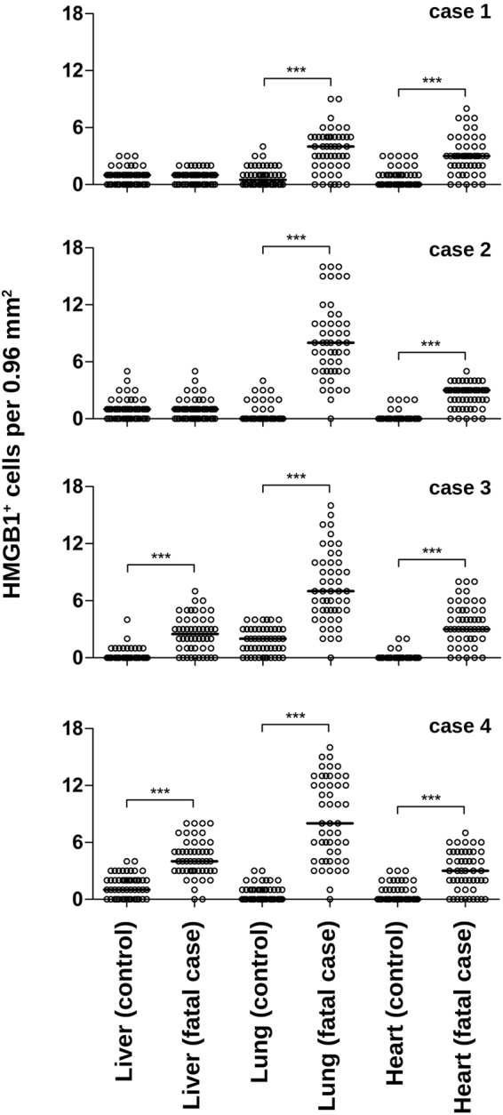
Figure 5.

Semi-quantitative analysis of HMGB1+ cells within post-mortem tissues of dengue fatal cases. For each studied organ (liver, lung and heart), 50 fields were acquired under a Nikon ECLIPSE E600 microscope coupled with a Cool SNAP-Procf Color camera. Cells with cytoplasmic HMGB1 expression were quantified using organ samples from four dengue fatal cases and four non-dengue control cases. Results are expressed as the median of HMGB1+ cells per 0.96 mm2. Mann-Whitney non-parametric statistical test was applied to verify the differences between groups (***p < 0.001).
