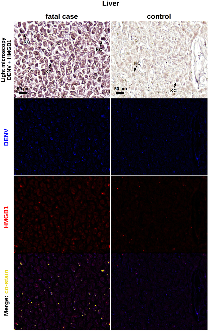
Figure 6.
Co-staining of DENV and HMGB1 in the liver. Liver sections from dengue fatal case (case 4) and a non-dengue case were processed for in situ hybridization and IHC assays. DENV was detected by a probe that anneals to a conserved sequence within the viral RNA negative strand and HMGB1 was assessed by immunohistochemistry assay. In the dengue case, the figure shows a diffuse area of hepatic steatosis with presence of DENV RNA in Kupffer cells (KC) and in sinusoidal borders of hepatocytes. Expression of HMGB1, when considering the dengue case, occurred within the nucleus as well as the cytoplasmic regions. DENV RNA and HMGB1 signals co-stained within the cytoplasm of hepatocytes and Kupffer cells. In the control case, KCs were negative for DENV and, in general, the HMGB1 expression was marginal and predominantly in nuclear regions.

