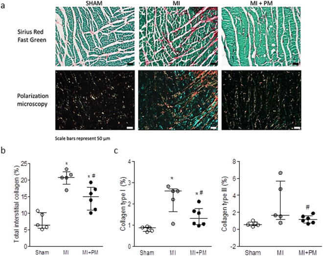
Figure 5.
Pyridoxamine reduces collagen content. (a, upper panel) Representative images of interstitial collagen obtained with Sirius red/Fast Green in the peri-infarct area. (a, lower panel) Representative images of collagen type I and type III, under polarized light microscopy. The red color indicates the highly cross-linked collagen type I, the green color indicates immature collagen type III. (b) Total interstitial collagen quantification in the peri-infarct area. (c) Collagen type I (left panel) and collagen type III (right panel) quantification in the peri-infarct region. Data are shown as median [75th percentile; 25th percentile] in Sham (N = 5), MI (N = 5) and MI + PM (N = 6). *Denotes p < 0.05 vs Sham, #denotes p < 0.05 vs MI.

