Figure 7.
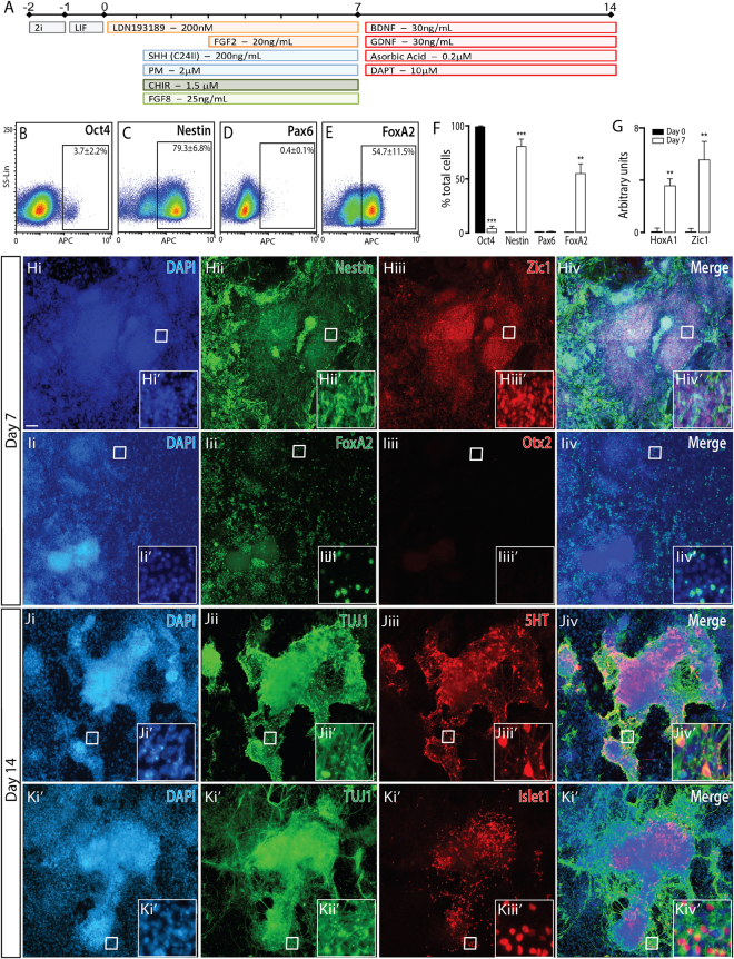
Naïve mESCs differentiated to a ventral hindbrain fate, show appropriate regional and temporal specification. (A) Schematic of the neural differentiation protocol, illustrating morphogen and small molecules employed, to generate ventral hindbrain progenitors and neurons from naïve PSCs. (B–E) Flow cytometry plots at day 7 and, (F) quantitative analysis at 0 and 7 for Oct4, Nestin, FoxA2 and Pax6, showing appropriate downregulaton of pluripotent (Oct4) and dorsal (Pax6) markers together with upregulation of neural (Nestin) and ventral (FoxA2) expression. (G) Transcriptional expression of hindbrain genes Hoxa1 and Zic1. (H) Representative photomicrographs depicting expression of DAPI, Nestin and Zic1 in hindbrain progenitors at day 7. (I) Ventral hindbrain specification of progenitors was confirmed by the presence of ventral marker FoxA2 and absence of mesodiencephalic protein Otx2. (J) By day 14, hindbrain specified cultures contained serotinergic neurons immunoreactive for TUJ1 and 5HT, as well as (K) Islet1+ TUJ1+ neurons, indicative of motor neurons. Data represents mean ± SEM, (n = 3), Students t-test. Scale bar = 100 um. **p < 0.01, ***p < 0.001.

