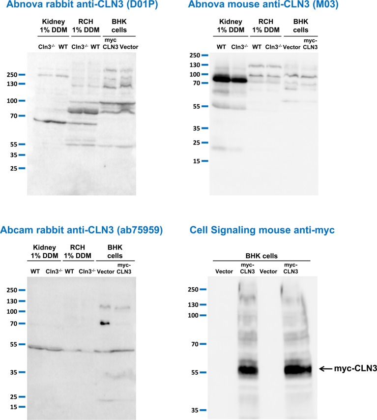
Figure 2. Anti-CLN3 antibodies do not detect CLN3 in mouse tissue extracts prepared with a special detergent or in BHK cells overexpressing human CLN3.
Three different anti-CLN3 antibodies [Abnova rabbit anti-CLN3 (D01P), Abnova mouse anti-CLN3 (M03), and Abcam rabbit anti-CLN3 (ab75959)] were tested in immunoblot experiments using protein extracts prepared with the nonionic detergent, n-dodecyl-β-D-maltopyranoside (DDM), from the kidney and the right cerebral hemisphere (RCH) of 254-day-old WT and 285-day-old Cln3−/− male mice. Extracts of BHK cells transfected with either a plasmid overexpressing myc-tagged human CLN3 or the plasmid vector only were also used in these experiments. BHK cell extracts were prepared with 1% DDM-containing lysis buffer. Sixty micrograms of protein were loaded in each lane of SDS-containing 10% polyacrylamide gels. Prior to loading, samples were incubated at 37°C for 30 min in reducing sample buffer containing 4 M urea. After the electrophoretic separation, proteins were transferred onto nitrocellulose membranes and probed with the anti-CLN3 antibodies (Abnova rabbit, 1:500; Abnova mouse, 1:500; Abcam rabbit, 1:700). The mouse anti-myc antibody from Cell Signaling was used in a dilution of 1:1000; M.W. marker: PageRuler Plus (Thermo Fisher Scientific). The immunoblots shown are representative of two separate experiments.

