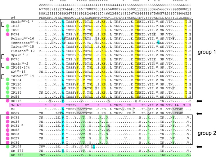
FIG 2.
Alignment of PBP2x sequences. Only sites of the transpeptidase domain that differ from PBP2x of S. pneumoniae R6 are shown. The vertical numbers in the first three rows indicate the amino acid positions. The asterisks above the amino acid position mark relevant mutations (see the text for details). The PBP2x variants of group 1 and group 2 are boxed. The color code for the mutations is as follows: light blue, mutations close to active-site motifs (aa 338, 394, and 550); yellow and green, signature mutations of group 1 and group 2 PBP2x, respectively; gray, mutations that occur only in particular variants. The reference sequences of S. mitis (Sm) strains M3 and 658 are shaded in pink and green, respectively. Black arrows on the right mark distinct PBP2x variants; brackets on the left indicate highly similar sequences. Green dots, strains from Iran; red dots, strains from Romania. 1, PBP2x of the Spain23F-1 clone is identical to PBP2x of the Spain9V-3 clone and S. pneumoniae CGSP14 (6); 2, a PBP2x variant identical to that in RO31 is present in RO58, RO61, and RO106. SA19A-13, South Africa19A-13 clone.

