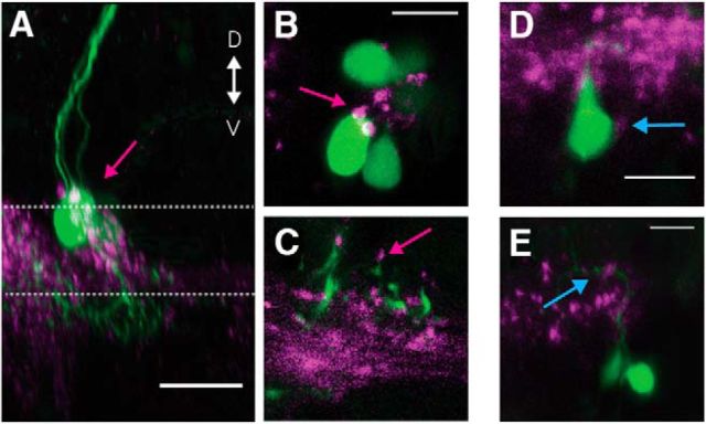
Figure 4.
Vestibular nucleus neurons show synaptophysin-positive puncta on their motoneuron targets. A, Sagittal MIP of a labeled SO motoneuron (magenta arrow) in green and the purple synaptic puncta labeled in Tg(−6.7FRhcrtR:gal4VP16); Tg(5×UAS:sypb-GCaMP3). Dotted lines indicate the planes in B, C. Scale bar, 20 μm. B, C, Close-up slice of the motoneuron somata in A with puncta (magenta arrow). Scale bar, 10 μm. D, Close-up of a retrogradely labeled SR motoneuron soma (green) with visible purple puncta (cyan arrow). Scale bar, 10 μm. E, Close-up of the dendrites of SR motoneurons (green) with visible purple puncta (cyan arrow). Scale bar, 10 μm.

