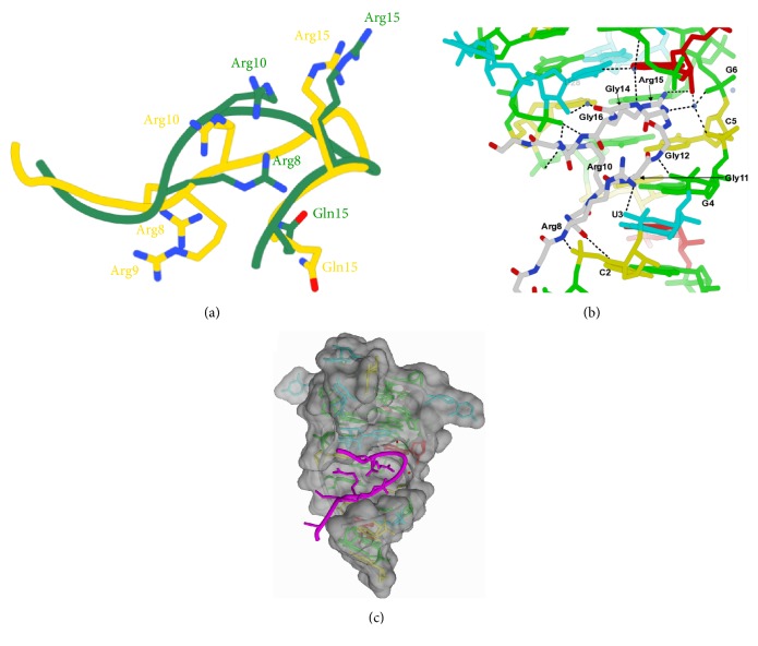
Figure 3.
RGGGGR peptide bound to SC1 RNA quadruplex-duplex junction. Comparison of peptide conformations between X-ray (green) and NMR (yellow) structures shown in (a). Hydrogen bonding pattern between peptide and nucleic acids shown in (b). Space filling model shown in (c), with peptide shown as a magenta ribbon. PDBs 5DEA (X-ray) and 2LA5 (NMR).

