
Figure 3.
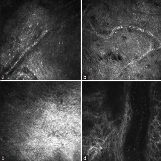
Corneal structure under confocal microscope at 28 days after modeling (400 μm × 400 μm): (a) Group A; (b) Group B; (c) Group C; (d) Group D. In the four groups, the matrix scar was observed, inflammatory cell infiltration was significantly reduced, and the corneal neovascularization formation was obvious. Blood cells flow within large vessels, especially common in Group D