
Figure 1.
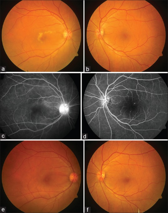
Serial fundus photography of the patient with dengue acute macular neuroretinopathy. (a) Colored fundus photograph of the right eye at baseline shows a grayish-yellow lesion with granular appearance and irregular borders surrounding fovea. (b) The left eye had the presence of a few hard exudates and one visible microaneurysm. (c) The late-phase angiogram of the right eye shows ill-defined faint hyperfluorescence in the perifoveal region along with disc staining. (d) The late arteriovenous phase angiogram of the left eye shows the presence of a few microaneurysms. (e) Colored fundus photograph of the right eye at 6-month follow-up shows near complete resolution of the grayish-white macular lesion, while the left eye shows some residual hard exudates with improvement in foveal transparency (f)