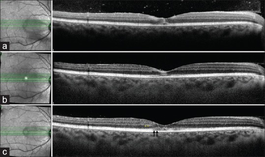
Figure 2.

(a) Optical coherence tomography line scan passing through the lesion in the right eye shows hyperreflectivity of the inner retinal layers (i.e. the ganglion cell layer, the outer plexiform layer, and the inner nuclear layer) in the parafoveal region along with hyperreflectivity of the outer retinal layers (outer plexiform layer, Henle's layer, and outer nuclear layer) at the fovea. In addition, there is disruption of the external limiting membrane, the ellipsoid zone, and the interdigitation zone in the foveal region. There is presence of hyperreflective dots in the preretinal space suggestive of vitreous cells. (b) The corresponding optical coherence tomography line scan at 4-week follow-up shows partial resolution of the hyperreflectivity of the inner retinal layers with restoration of the foveal contour. There is only partial restoration of the external limiting membrane, ellipsoid zone, and interdigitation zone layers. (c) The optical coherence tomography line scan at 6-month follow-up shows further resolution of the inner and outer layer hyperreflectivity with better delineation of the individual layers. However, there is mild residual disruption of the external limiting membrane, ellipsoid zone, and interdigitation zone layers (black arrows) with thinning of the outer nuclear layer (yellow asterisk). There is complete resolution of the overlying vitritis
