
Figure 2.
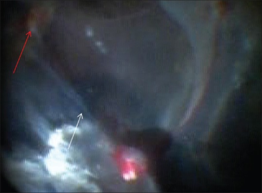
Intraoperative fundus photograph of the left eye showing proliferative retinopathy with extensive tractional retinal detachment (white arrow) involving the posterior pole. Red arrow shows the position of the optic nerve head
Official websites use .gov
A
.gov website belongs to an official
government organization in the United States.
Secure .gov websites use HTTPS
A lock (
) or https:// means you've safely
connected to the .gov website. Share sensitive
information only on official, secure websites.
Intraoperative fundus photograph of the left eye showing proliferative retinopathy with extensive tractional retinal detachment (white arrow) involving the posterior pole. Red arrow shows the position of the optic nerve head